Age: 61 years Gender: Male Site: Lower lip Duration: 3 months Morphology: An asymptomatic, solitary, dome-shaped nodule with keratotic surface. Excisional biopsy revealed KA.
Age: 80 years Gender: Female Site: Nose Duration: 1 year History: A small persistent crusted erosion with with no tendency for spontaneous healing or resolution. Excisional biopsy confirmed BCC.
Age: 50 years Gender: Male Site: Lower lip Duration: 3 months Morphology: An asymptomatic, solitary, well-circumscribed, dome-shaped, pinkish, crusted nodule about one cm diameter. Excisional biopsy showed KA.
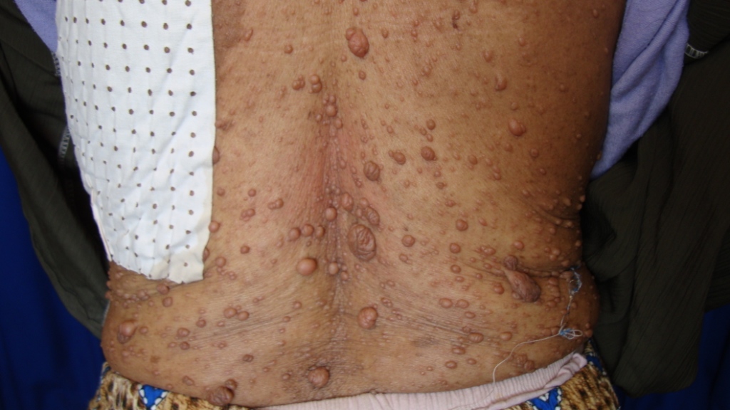
Age: 38 years Gender: Female Site: Back Duration: 20 years Morphology: Myriad of asymptomatic, flesh-colored, soft nodules with buttonholing sign. These begnin skin tumors were associated with multiple cafe-au-lait patches.
Age: 46 years Gender: Female Site: Leg Duration: 9 months History: An asymptomatic, solitary, indurated and infiltrative crusted plaque having an inverted-heart shape with more activity on the marginal zone than the center.
Age: 78 years Gender: Male Site: Scalp Duration: 1 year History: 11 years ago this man had BCC on his scalp removed surgically. Now he presents with a noduloulcerative lesion with beefy red base and pigmented rolled up border at the same site of previous lesion. Biopsy confirmed BCC.
Age: 69 years Gender: Male Site: Face & Trunk Duration: > 2 years History: Asymptomatic, firm to hard, infiltrative, dull red plaques and nodules. Lymph nodes enlargement and hepatomegally were positive.
Age: 20 years Gender: Female Site: Face Duration: Since puberty Morphology: Asymptomatic, multiple, firm, translucent papules involved the midface especially the nasolabial folds.
Age: 68 years Gender: Male Site: Scalp Duration: Recurrent (Decades) History: At 1950s the patient was treated with radiation therapy for favus (the treatment available for tinea capitis at that time). Many decades later (1990s) he developed BCC on his atrophic scalp. This time it was treated with surgical excision. Three years later it recurred and treated with an unwise decision with radiation therapy (10 sessions ). Now he presented with a large pigmented plaque having slightly raised border.
Age: 66 years Gender: Female Site: Nose Duration: 5 years Morphology: Multiple BCCs involving the face of this woman with xeroderma pigmentosum. The main one was phagedenic ulcer with characteristic rolled up border. It has destroyed most of the nose and exposed its interior.